
Pulmonary Embolism Nursing Care
 By Maureen Osuna, MSN, RN on December 19, 2024
On mobile? Review pulmonary embolism nursing in episode 378 from your favorite podcast player here.
Follow the show! If you loved this episode, be sure to follow the show in your favorite podcast app so you never miss an episode…and share with friends so you can help other nurses thrive!
What is a pulmonary embolism?
In this article, you’ll learn the key things you need to know about pulmonary embolism nursing care. A pulmonary embolism is a condition in which vessels in the lungs are blocked by a thrombus or other material that has traveled from somewhere else in the body. This is usually a result of a venous thromboembolism, but can also be due to amniotic fluid, air or fat embolus, or tumor material in patients with cancer.
A key thing to understand about pulmonary embolism is that the airway is not occluded. In pulmonary embolism, one or more blood vessels are occluded. What this means is that blood coming from the right side of the heart isn’t able to pass freely through the pulmonary vasculature to participate in gas exchange, which negatively affects perfusion.
How serious is pulmonary embolism?
Pulmonary embolism caused by venous thromboembolism (VTE) is a leading cause of preventable hospital death in the United States. This is why so much emphasis is placed on VTE prophylaxis in the clinical setting. This includes compression stockings, sequential compression devices (SCDs) and anticoagulants. To learn more about VTE prevention, check out this article on deep vein thrombosis.
Left untreated, acute pulmonary embolism has a mortality rate as high as 30%, and up to 10% of patients die suddenly. However, with prompt treatments, mortality rates can be as low as 8%. In order for patients to have the best possible outcome, pulmonary embolism must be recognized and treated quickly.
Who is at risk for pulmonary embolism?
A key component of quick recognition is knowing who is at risk for pulmonary embolism. In most cases, this is a patient who is at risk for developing a VTE. This includes patients who are immobile, those who have had recent surgery or trauma (such as a leg fracture), and those with hypercoagulability (such as patients taking estrogen or patients with elevated platelet counts).
It’s important to note that PE is not always due to VTE. Other causes include:
- Fat embolism – This occurs when fat from a long bone or pelvic bone enters systemic circulation as a consequence of surgery or trauma.
- Amniotic fluid embolism – This occurs when amniotic fluid, fetal cells or other debris enter maternal pulmonary circulation. Learn more about this complication of pregnancy here.
- Tumor fragments – Though not common, tumor fragments or clusters of tumor cells can dislodge and travel to the pulmonary vasculature in patients who have solid tumor cancer.
- Air embolism – This complication results when air is introduced into systemic circulation. This can occur during surgery, scuba diving, trauma, and placement and removal of central venous catheters.
Pulmonary embolism pathophysiology
- When a thrombus occludes pulmonary circulation, catecholamines and inflammatory mediators are released. These neurohormonal substances cause vasoconstriction and further impede blood flow to the lung.
- This vasoconstriction causes increased pulmonary pressure, which causes the right side of the heart to have to work harder. This can lead to heart failure and reduced cardiac output.
- When a portion of the lung does not get blood flow, this leads to damaged lung tissue and a loss of surfactant. This results in a V/Q mismatch, also known as a ventilation-perfusion mismatch. The short explanation is that when a patient has ventilation but inadequate perfusion, this is a type of V/Q mismatch called a “dead-space” mismatch. The air is getting to the alveoli, but the alveoli are not able to participate in gas exchange due to the presence of the thrombus, which is impeding blood flow. Take a deep dive into V/Q mismatch here!
- Overall, the patient experiencing a pulmonary embolism can have significant hypoxia, reduced cardiac output, and cardiac dysrhythmias. In severe cases, they can go into shock, which has a high mortality rate.

Now that you’ve got some background information on pulmonary embolisms, let’s learn about how to care for these patients using the Straight A Nursing LATTE method.
L: How does the patient LOOK?
A patient experiencing pulmonary embolism is likely to have the following signs and symptoms:
- Shortness of breath (can be significant!)
- Dropping Spo2
- Increased work of breathing
- Chest pain upon inspiration and/ or chest pressure
- Cough, with or without blood-tinged sputum
- Anxiety
- Feeling of impending doom
A: What ASSESSMENTS are part of pulmonary embolism nursing care?
- Obtain a set of VS, which can show low SpO2, tachypnea, hypotension and tachycardia. Want to take a deep dive into how a pulmonary embolism can cause hypotension? Click this link to learn more!
- Assess work of breathing and for signs of air hunger such as gasping or grunting which are signs of severe respiratory failure.
- Listen to the lungs which may reveal coarse lung sounds or a pleuritic friction rub.
- Listen to the heart sounds, S3 and S4 may be present in severely compromised patients.
- Assess for cyanosis, which is a bluish discoloration of the skin. In darker skinned individuals, cyanosis may appear more gray-ish or pale.
- Assess for confusion or decreased LOC which occur when the brain is not getting adequately perfused.
- Assess the skin for petechiae which are small reddish dots under the skin – if present, these occur on the chest and axilla. These are most likely to occur if the PE is due to a fat embolism.
- Monitor for signs of bleeding secondary to thrombolytic therapy which is discussed in the treatment section below.

T: What tests will be utilized to confirm or evaluate pulmonary embolism?
- ABG – An arterial blood gas will reveal low oxygen levels and initially low PaCO2 due to hyperventilation. As the condition progresses, the CO2 level will rise leading to respiratory acidosis.
- D-Dimer – In the case of a blood clot, the D-Dimer will be elevated. This indicates the release of fibrin degradation products in response to clot formation.
- Chest x-ray – Though the fastest of the imaging studies used to evaluate pulmonary embolism, an x-ray is likely only going to show larger embolisms.
- Chest CT scan – A CT scan is relatively quick and is one of the most commonly utilized diagnostic exams for patients suspected of having a pulmonary embolism.
- V/Q scan – A ventilation-perfusion scan looks at the circulation of blood in the pulmonary vasculature. While this test is highly effective in diagnosing pulmonary embolism, it takes much longer to perform than a CT scan. In addition, the patient has to lie flat for an extended period of time which can be incredibly difficult when shortness of breath is severe.
- Pulmonary angiography – This exam is highly diagnostic for PE, but invasive and expensive. In this exam, the physician inserts a long, thin catheter through a vessel and advances it all the way into the pulmonary artery. Dye is then injected into the catheter which enables the physician to take x-ray images that can reveal any blockages in pulmonary vasculature.
- Other imaging studies – Ultrasound or MRI may be used to help locate the embolism, particularly in situations where CT is not available.
- EKGs – A 12-lead EKG will typically reveal tachycardia and could also show the S1Q3T3 pattern. This looks like a large S wave in lead I along with a Q wave and inverted T wave in lead III. This pattern is indicative of strain on the right side of the heart.
- Coags – Coagulation labs will be drawn prior to initiating thrombolytic treatment and regularly throughout therapy.
- Fibrinogen – If receiving TPA, the patient will need fibrinogen evaluated routinely.
- BNP – In cases where heart failure is suspected, blood will be drawn to test the level of brain natriuretic peptide (it is elevated in heart failure).
T: What TREATMENTS are provided for pulmonary embolism?
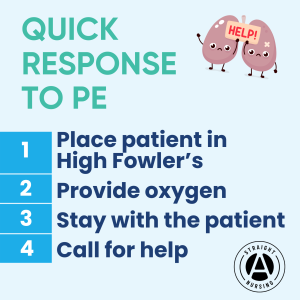
Initial nursing interventions for someone suspected of a PE are to place the patient in high-fowler’s position and supply supplemental oxygen per protocol. Other treatments include:
Thrombolytics – Thrombolytics are medications used to break up an existing clot (ex: TPA).
Anticoagulants – Anticoagulants may also be utilized to prevent future clots from forming. The most commonly utilized anticoagulant is unfractionated heparin, though you may see enoxaparin as well. Patients may also need to take long-term anticoagulation, which is often warfarin or a direct oral anticoagulant (DOAC) such as apixaban (Eliquis).
IVC filter – Patients who are at high risk for PE due to VTE may get an inferior vena cava filter placed. This filter essentially “catches” and breaks up any emboli as they travel from the deep veins of the lower body up through the vena cava.
Embolectomy – Larger clots may require a surgical procedure called an embolectomy.
Other treatments – Patients with reduced cardiac output may need fluids, inotropic medications (such as dobutamine or milrinone) and others may need vasodilators to combat pulmonary hypertension.
E: How do you EDUCATE the patient about pulmonary embolism?
For a patient receiving anticoagulation:
- Teach the patient/family about bleeding precautions which includes things like shaving with electric razors and being extremely careful when using sharp instruments such as scissors or knives. They will likely be advised to avoid activities that put them at high risk for falls or injury such as contact sports and even cycling.
- Teach the patient and family to seek medical attention with any head injuries due to the heightened risk for bleeding.
- Ensure the patient understands they may bruise easily, and to report any increased bruising to their MD as this could indicate they have been overly anticoagulated.
- Have the patient or caregiver explain to you how they will take their medication and any labs that will be routinely monitored such as INR monitoring with warfarin.
- If the patient is taking warfarin, teach them that they will need to either avoid foods high in vitamin K or have a consistent amount of foods high in vitamin K. The most common source of dietary Vitamin K is leafy greens, which can counteract therapy.
Also, teach your patient that they are at higher risk for recurrent blood clots. They should be instructed to seek medical attention if any symptoms of blood clot or pulmonary embolism return. In addition, regular self checks of the extremities should be completed, looking for any areas of hardness, swelling, or pain, which are indicators of clot formation.

Looking for more articles covering respiratory conditions? Check out this collection here!
_____________________________________
The information, including but not limited to, audio, video, text, and graphics contained on this website are for educational purposes only. No content on this website is intended to guide nursing practice and does not supersede any individual healthcare provider’s scope of practice or any nursing school curriculum. Additionally, no content on this website is intended to be a substitute for professional medical advice, diagnosis or treatment.
References:
Bĕlohlávek, J., Dytrych, V., & Linhart, A. (2013). Pulmonary embolism, part I: Epidemiology, risk factors and risk stratification, pathophysiology, clinical presentation, diagnosis and nonthrombotic pulmonary embolism. Experimental & Clinical Cardiology, 18(2), 129. https://pmc.ncbi.nlm.nih.gov/articles/PMC3718593/
Lippi, G., Salvagno, G. L., Ippolito, L., Franchini, M., & Favaloro, E. J. (2010). Shortened activated partial thromboplastin time: causes and management. Blood Coagulation & Fibrinolysis: An International Journal in Haemostasis and Thrombosis, 21(5), 459–463.
Lome, S. (n.d.). Pulmonary Embolism ECG Review. Healio. https://www.healio.com/cardiology/learn-the-heart/ecg-review/ecg-topic-reviews-and-criteria/pulmonary-embolism
Mayo Clinic. (n.d.). Pulmonary embolism – Symptoms and causes. Mayo Clinic. https://www.mayoclinic.org/diseases-conditions/pulmonary-embolism/symptoms-causes/syc-20354647
McRae, S., & Ginsberg, J. (2005). The diagnostic evaluation of pulmonary embolism. 3, 14–20. https://doi.org/10.1111/j.1541-9215.2005.03698.x
Medline Plus. (2022). Pulmonary angiography. MedlinePlus Medical Encyclopedia. https://medlineplus.gov/ency/article/003813.htm
Newman, T. (2023, June). Air embolism: Causes, symptoms and treatment. https://www.medicalnewstoday.com/articles/186328
Ouellette, D. (n.d.). Pulmonary Embolism (PE): Practice Essentials, Background, Anatomy. https://emedicine.medscape.com/article/300901-overview
Reddy, N. M., Hall, S. W., & MacKintosh, F. R. (1999). Partial Thromboplastin Time: Prediction of Adverse Events and Poor Prognosis by Low Abnormal Values. Archives of Internal Medicine, 159(22), 2706–2710. https://doi.org/10.1001/archinte.159.22.2706
Rosovsky, R., Davis, G. A., & Smith, R. V. (2019, July). DOACs: Oral Anticoagulant Treatment of Choice for Pulmonary Embolism? Endovascular Today. https://evtoday.com/articles/2019-july-supplement/doacs-oral-anticoagulant-treatment-of-choice-for-pulmonary-embolism
Tarbox, A. K., & Swaroop, M. (n.d.). Pulmonary embolism Tarbox AK, Swaroop M – Int J Crit Illn Inj Sci. https://www.ijciis.org/article.asp?issn=2229-5151;year=2013;volume=3;issue=1;spage=69;epage=72;aulast=Tarbox
Last Updated on December 19, 2024 by Maureen Osuna, MSN, RN

share this
20 Secrets of Successful Nursing Students
Being a successful nursing student is more than just study tips and test strategies. It’s a way of life.


